What Are the Two Main Families of Parasitic Ticks Small Animal Care and Manaagement
Amblyomma americanum and Amblyomma spp.
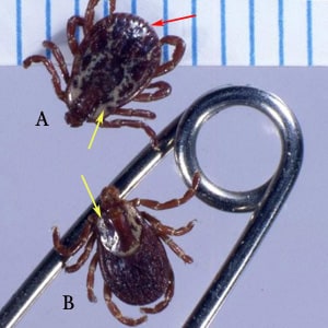
Members of the genus, Amblyomma, are known vectors of a variety of diseases in humans. In N America, A. americanum transmits Francisella tularensis (tularemia), Ehrlichia chaffensis (ehrlichiosis), and Rickettsia rickettsii (Rocky Mountain spotted fever, or RMSF). In Africa, A. hebraeum transmits Rickettsia conorii (boutonneuse fever); in Primal and South America, A. cajennense transmits RMSF. Members of the genus Amblyomma are characterized past having mouthparts noticeably longer than the basis capituli, a usually ornate dorsal shield, eyes present on the dorsal shield, and festoons (which may exist difficult to see in engorged specimens). Adult females of A. americanum accept a distinctive white spot near the posterior cease of the dorsal shield.
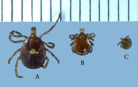
Figure A: Adult female (A), male (B), and nymph (C) of A. americanum. Notice the characteristic white spot on the female person's dorsal shield. Image courtesy of James Occi.
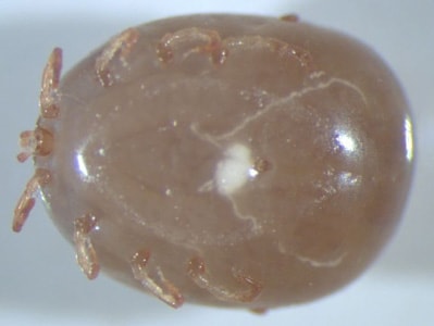
Figure E: Ventral view of an engorged nymph of Amblyomma sp., collected on a patient with travel history to Peru. Notice the festoons are non visible in the specimen due to the engorged state. Images courtesy of the Washington Country Public Health Laboratories.
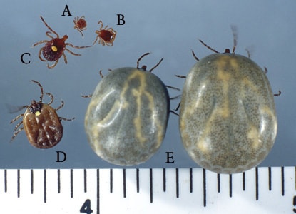
Figure B: Nymph (A), male (B), female (C), partially-engorged female (D) and two fully-engorged adults (E) of A. americanum. Image courtesy of James Occi.

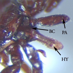
Effigy C: Close-upward of the mouthparts of A. americanum. Find the mouthparts (MP) and palps (PA) are long, in comparing with the basis capituli (BC).
Figure D: Dorsal view of an engorged nymph of Amblyomma sp., collected on a patient with travel history to Republic of peru. Notice the festoons are not visible in the specimen due to the engorged state. Images courtesy of the Washington Land Public Wellness Laboratories.
Dermacentor andersoni and D. variabilis.
Members of the genus, Dermacentor, are known vectors of Rickettsia rickettsii (Rocky Mount spotted fever, or RMSF), Colorado tick fever virus, Francisella tularensis (tularemia), Rickettsia sibirica (Siberian tick typhus), and Central European tick-borne encephalitis virus. They have also been implicated in tick paralysis. In Due north America, the ii well-nigh important species medically are D. variabilis and D. andersoni, the latter of which is more than prevalent in the Pacific Northwest and the former in the southward and eastward. Even so, their distributions may overlap in the west, and identification to the species level is best obtained past comparing the morphologic features of the spiracular plates. Developed members of the genus Dermacentor are characterized by a ordinarily ornate dorsal shield, mouthparts brusque (in comparing with the basis capituli), eyes on the dorsal shield, and the presence of festoons (which may be difficult to run into in engorged specimens).
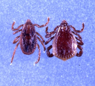
Effigy A: Male (A) and female (B) of D. variabilis. Observe the ornate dorsal shields (yellow arrows), which on the male covers nigh of the tick's body. Also find the presence of festoons (cherry arrow). Epitome courtesy of James Occi.

Effigy E: Spiracular plate of D. variabilis. Notice the slight dorsal prolongation (arrow) of the plate and large number of smaller goblet cells on the plate.
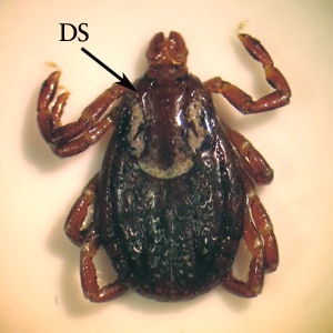
Effigy B: Female specimen of D. variabilis. Notice the ornate dorsal shield (DS).

Effigy F: Spiracular plate of D. andersoni. Notice the more pronounced dorsal prolongation (arrow) of the plate and the larger, yet fewer in number, goblet cells on the plate. Paradigm courtesy of the Washington Land Public Health Laboratories.
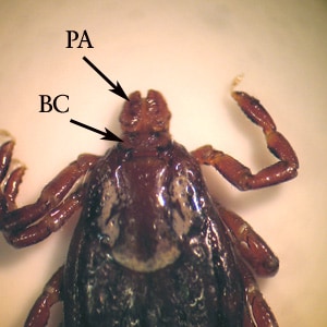
Figure C: Higher-magnification of the specimen in Effigy B. Notice the palps (PA) are brusk in relation to the basis capituli (BC).
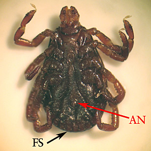
Figure D: Underside of the specimen in Figure B, showing the anus (AN) and festoons (FS).
Ixodes scapularis and Ixodes spp.
Members of the genus, Ixodes, are known vectors of Borrelia burgdorferi (Lyme disease), Babesia spp. (babesiosis), human granulocytic ehrlichiosis (HGE), and Russian spring-summer encephalitis virus. In N America, the two most of import species medically are I. scapularis and I. pacificus. Adults are characterized by having mouthparts longer than the ground capituli, a lack of festoons, an inornate dorsal shield without eyes, and an inverted, U-shaped anal groove.
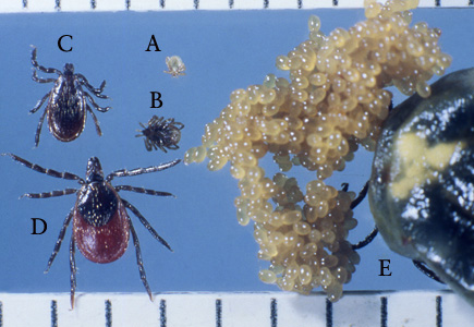
Figure A: Larva (A), nymph (B), adult male (C), adult female (D), and engorged adult female with eggs (E) of I. scapularis. Image courtesy of James Occi.
Effigy E: Dorsal view of the specimen in Figure C, showing a close-upwards of the inductive region. Discover the hypostome (HY) and palps (PA) are long, in relation to the basis capituli (BC).
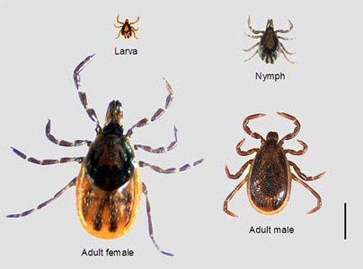
Figure B: Adult female, adult male, nymph and larva of I. scapularis. Image courtesy of Dr. Marc Dolan.
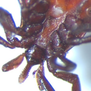
Figure F: Ventral view of the specimen in Figure C, showing a shut-upward of the anterior region.
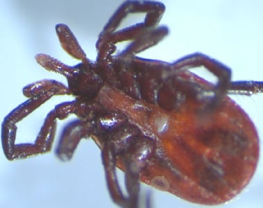
Figure C: Ventral view of a nymph of Ixodes sp. Image courtesy of the Washington Country Public Health Laboratories.
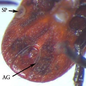
Figure D: Higher-magnification of the specimen in Figure C, showing a spiracular plate (SP) and the inverted, U-shaped anal groove (AG). Discover besides the absence of festoons.
Rhipicephalus sanguineus.
Rhipicephalus sanguineus is known as the brown dog tick and is found about worldwide. Humans are not the usual host, only there are increasing reports of illness transmission with this species, including Rickettsia rickettsii (Rocky Mountain spotted fever, or RMSF) and Rickettsia conorii (boutonneuse fever). Adults are characterized by having a laterally-produced, angulate basis capituli, a dorsal shield with eyes, festoons, and deeply-cleft forepart coxae. The festoons and anal groove may be difficult to run into in engorged specimens. The mouthparts are relatively short, in relation to the basis capituli.

Figure A: Female of R. sanguineus.
Effigy E: Female (left) and male (right) of R. sanguineus. Image courtesy of James Occi.
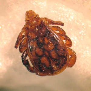
Figure B: Male person of R. sanguineus.
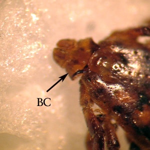
Figure C: Close-upward of the specimen in Figure B. Discover the laterally-produced, angulate basis capituli (BC).
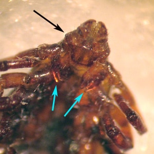
Figure D: Ventral view of the specimen in Figures B and C. Discover the laterally-produced, angulate basis capituli (black arrow) and deeply-cleft fore coxae (blue arrows).
Ornithodoros moubata and O. turicata.
Members of the genus, Ornithodoros, are known vectors of Borrelia hermsi (tick-borne relapsing fever, or TBRF) in N America and several TBRF spirochetes in Africa. Like about members of the Argasidae, or soft ticks, Ornithodoros spp. are characterized by lacking a dorsal shield and not displaying marked sexual dimorphism. Their mouthparts are subterminally attached and non visible from above. Adults and developing nymphs do not remain attached to their hosts, as do members of Ixodidae; rather they are adapted to feeding rapidly and leaving the host promptly.
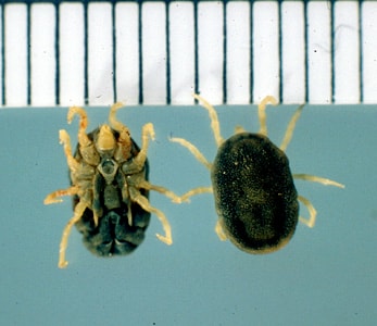
Figure A: Ventral (left) and dorsal (right) views of O. turicata, a known vector of TBRF spirochetes in the midwestern and southwestern The states. Image courtesy of James Occi.

Effigy B: Ornithodoros moubata, a known vector for African TBRF spirochetes. Image courtesy of James Occi.
Source: https://www.cdc.gov/dpdx/ticks/index.html
0 Response to "What Are the Two Main Families of Parasitic Ticks Small Animal Care and Manaagement"
Post a Comment